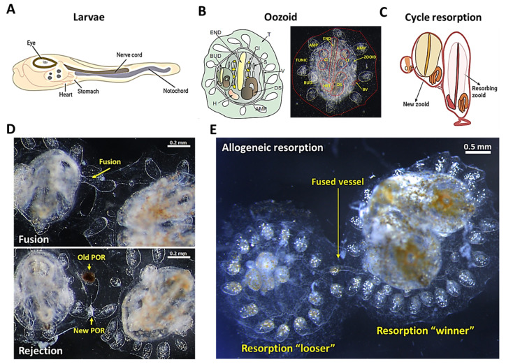
Figure 1.
The anatomy of B. schlosseri and its different levels of naturally occurring immune responses. (A) Diagram of the tunicate larvae tadpole phase showing the nerve cord and the notochord. (B) Diagram and live imaging of the ventral view of a zooid (Z) and primary bud (BUD), embedded within a tunic (TUN) and connected with vasculature (V), which terminates in ampullae (AMP). The zooid has a branchial sac conformed by the endostyle (END), stigmata (S), cell islands (CI), digestive system (DS), and heart (H). (C) Diagram showing the “takeover” phase in the weekly cycle of zooid regeneration mediated by noninflammatory programmed cell removal of the resorbing old zooid. (D) Live imaging of two B. schlosseri colonies undergoing fusion (arrows show fused vasculature (top)) and rejection (arrows show points of rejection (POR) (bottom)). (E) Live imaging from the allogeneic resorption process, where one colony is the “loser” (which is resorbed), while the other is the “winner”, demonstrating normal developmental stages. (A,C) were created using BioRender; (B,D) were reproduced with permission from [17], Springer Nature Limited, Berlin, Germany, 2018; (E) was reproduced with permission from [16], National Academy of Sciences, 2016.

