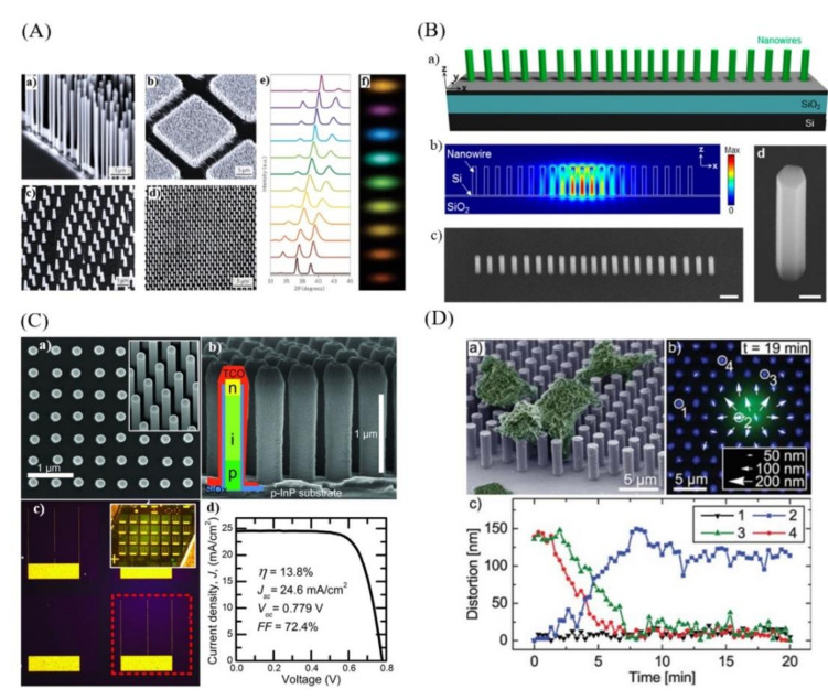
Figure 1.
(A) Scanning-electron microscope images of different vertically aligned semiconductor nanowire arrays (ZnO NWs (a), GaN NWs (b), InP NW (c), InAs/InP heterostructured NWs (d)), (e) X-ray diffraction for InGaN nanowires arrays with different In concentrations, (f) CCD camera image of the visible photoluminescence emission of InxGa1−xN nanowire (x = 0–0.6) (reprinted with permission of Refs. [20,76,77,78]); (B) InGaAs/InGaP core/shell nanowires array laser monolithically integrated on SOI: (a) Schematic of the nanowire array laser, (b) Electric field profile (|E|) of the fundamental cavity mode, (c) Tilted SEM images of the nanowire array laser and (d) individual NW (reprinted with permission of Ref. [19]); (C) InP nanowire array solar cells: (a) top-view and 30° tilt scanning electron micrographs of as-grown NWs, (b) SEM images of processed nanowires with a schematic of the individual devices, (c) optical microscope images of the solar cells, (d) solar cell I-V characteristics (reprinted with permission of Ref. [16]); (D) NWs array based nanomechanical biosensor for living cells induced forces detection: (a) scanning electron micrograph of the cells immobilized on the array, (b) fluorescence of the cell and reflection of the nanowire array, (c) distortion analysis as a function of time. (Reprinted with permission of Ref. [81]).

