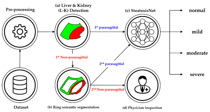
Figure 3.
Simplified flowchart of the proposed method: (a) Liver and kidney (L-K) detection yielding 1st parasagittal and non-parasagittal images. (b) Ring detection to double-check the 1st non-parasagittal image, producing 2nd parasagittal and non-parasagittal images. (c) Grading of the 1st and 2nd parasagittal images using SteatosisNet according to the level of steatosis. (d) Grading the steatosis level of the 2nd non-parasagittal images by physician inspection.

