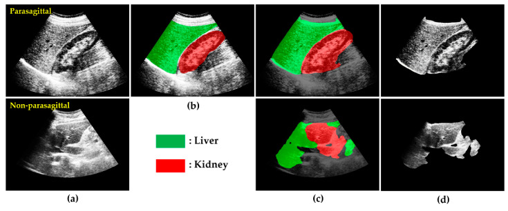
Figure 5.
Some results of L-K semantic segmentation and L-K image cropping: (a) HE images. (b) Ground truth of US image with L-K area labeled. (c) L-K labeled images overlaid onto those in (a), with liver highlighted in green and kidney in red. (d) images. Top row: parasagittal; Bottom row: non-parasagittal.

