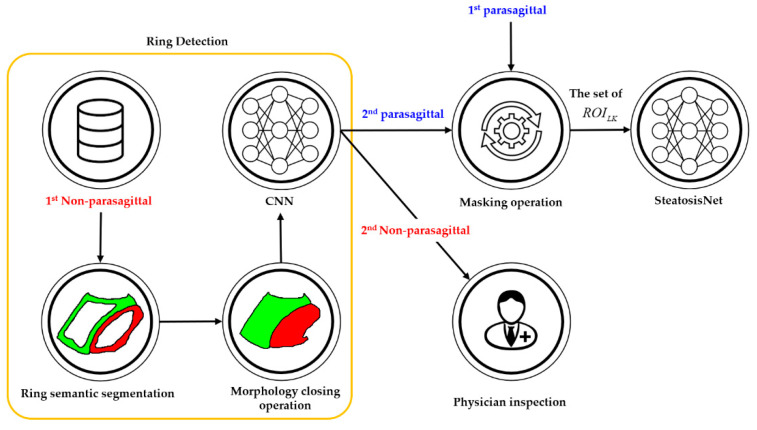
Figure 8.
Simplified flowchart of ring detection, where physicians can inspect the 2nd non-parasagittal images to grade the steatosis level. Ring segmentation produced a ring-segmented image, and the morphology closing operation yielded an L-K labeled image. The set of ROILK images was inputted to SteatosisNet.

