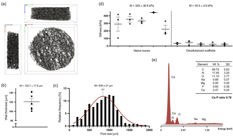
Figure 1.
Structural characterization of human femoral head-derived decellularized bone scaffolds. (a) A 3D volume rendering obtained from mesh segmentation (106 tetrahedra elements) of microCT scan of a representative scaffold. Box dimension: 10 × 10 × 3 mm. Axis: x—green, y—red, z—blue. (b) Wall thickness, or trabeculae thickness (Tb.Th), calculated from the total volume of interest (VOI) determined on SimplewareTM ScanIP respectively for each segmented mesh sample (n = 7). (c) Histogram of the relative frequency of pore size diameter (n = 5). Data assume a Gaussian distribution shown in red. (d) Stiffness of native and decellularized constructs, shown as means of elastic modulus obtained by mechanical compression testing (n = 5). The same shape and size were used for measurements of both native and decellularized samples. (e) Representative EDS spectra of decellularized scaffolds surface and respective quantification of atomic element weight percentage (wt%, n = 2). C—carbon, N—nitrogen, O—oxygen, Na—sodium, Mg—magnesium, P—phosphorus, and Ca—calcium.

