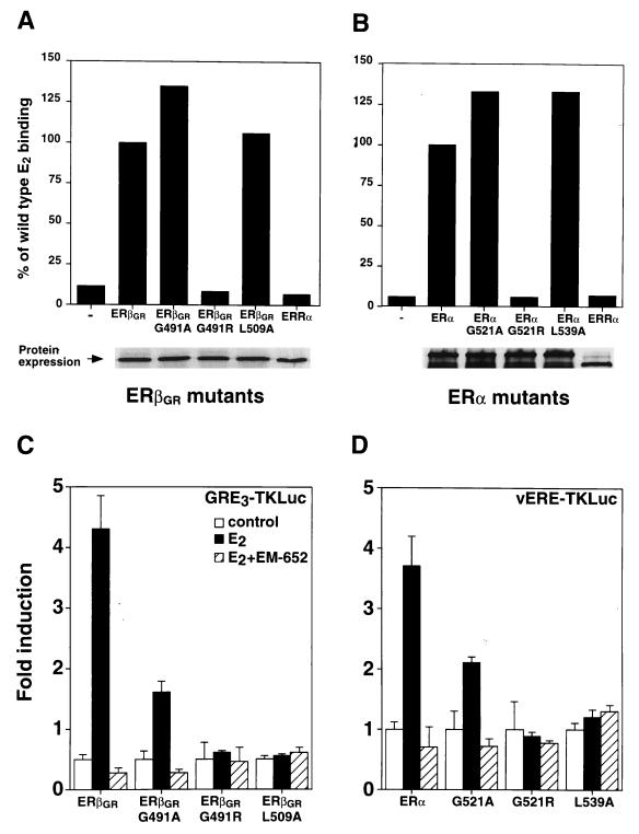
FIG. 6.
Functional characterization of ERβGR and ERα LBD mutants. (A) E2 binding analysis of in vitro-translated ERβGR mutants. Controls were conducted by using either unprogrammed reticulocyte lysates (−) or human ERRα, both of which are unable to bind E2. Results are expressed as the percentage of ERβGR ligand binding, which was arbitrarily set at 100%. The bottom panel shows that all receptors were expressed in equal amounts by 35S-labeling in parallel reactions. (B) Conditions were identical to those for panel A except that an analysis of the corresponding ERα mutants was conducted. (C) Cos-1 cells were cotransfected with the GRE3TKLuc reporter construct and wild-type or mutated ERβGR. The cells were treated with a control (0.1% ethanol) or 10 nM E2 in the absence or presence of 100 nM EM-652. (D) Conditions were identical to those for panel C except that Cos-1 cells were cotransfected with ERα receptors and the vERE1TKLuc reporter construct.

