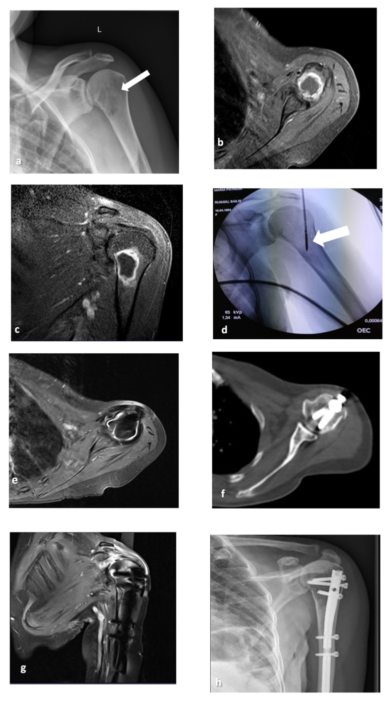
Figure 2.
A 56-year-old female patient with a left humerus metastasis from breast cancer (a–d). Coronal X-ray shows a lytic lesion in the superior portion of the humerus (arrow). Axial and coronal T1-weighted MR scans with fat suppression after gadolinium infusion (b,c) show the lytic lesion surrounded by a rim of enhancement. The coronal C-Arm image (d) shows the MW antenna positioned in the lesion (arrow). After 1 year of follow-up, axial and coronal MRI (e–g), axial CT (f) and X-ray (h) show the presence of the endoprosthetic replacement and no local relapse in the treated site.

