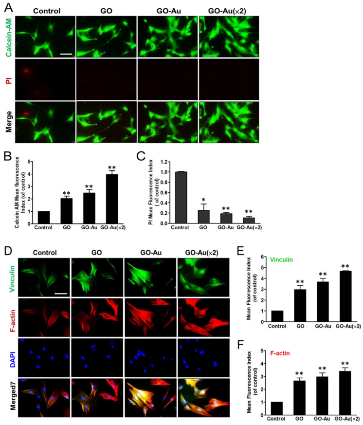
Figure 2.
Effects of different materials on MSCs viability using calcein-AM/PI co-staining assay. Cells were cultured in cover slide deposited with nanomaterials including GO, GO-Au, and GO-Au (×2) for 24 h. Cells were, then, co-stained with calcein-AM dye and PI, and cell viability was observed using a fluorescence microscopy. (A) Both the live cells and dead cells were presented in green and red fluorescence images, respectively. The fluorescence intensity in either (B) calcein-AM (live cells) or (C) PI (dead cells) with further quantification and analysis using Image J software. The control group is indicated as a glass cover slide. Scale bars, 20 µm. Data were presented as the mean ± SD, * p < 0.05 and ** p < 0.001. Effects of different materials on MSCs adhesion ability and cell morphology: (D) After cells were cultured on various nanomaterials of GO, GO-Au, and GO-Au (×2) for 24 h, then, cells were analyzed for cell adhesion capacity using an immunofluorescence assay and recognized with anti-vinculin antibody (green) and cytoskeletal F-actin phalloidin dye (red). Nuclei were stained with DAPI (blue). The representative fluorescent intensity of (E) vinculin and (F) F-actin were determined from three independent results. Scale bar, 20 μm. Data were presented as the mean ± SD, ** p < 0.001.

