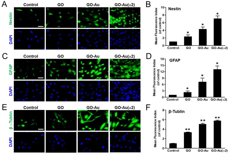
Figure 6.
Characterization of GO-Au-mediated MSC differentiation into neuron cells. Cells were cultured on various nanomaterials of GO, GO-Au, and GO-Au (×2) for 7 days. Then, cells were analyzed for expression levels of (A,B) nestin, (C,D) GFAP and (E,F) β-tubulin, using an immunofluorescence assay. Scale bar, 20 μm. The quantification of fluorescent intensity in nestin, GFAP, and β-tubulin protein expression. Data were presented as the mean ± SD, * p < 0.05 and ** p < 0.01.

