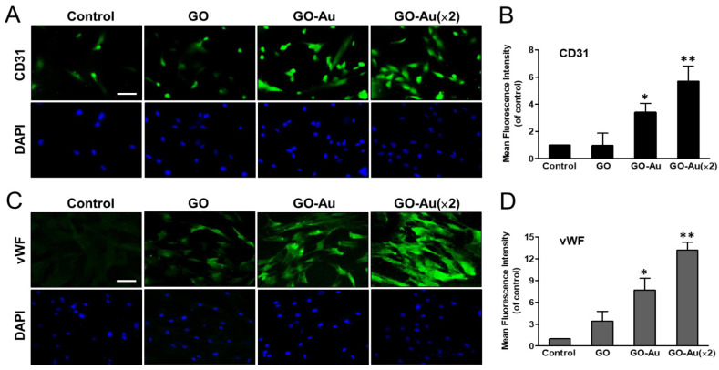
Figure 7.
Identification of GO-Au-mediated MSC differentiation into endothelial cells. Cells were cultured on various nanomaterials of GO, GO-Au, and GO-Au (×2) for 7 days. Then, cells were analyzed for the expression levels of (A,B) CD31 and (C,D) vWF, using an immunofluorescence assay. Scale bar, 20 μm. The quantification of fluorescent intensity in CD31 and vWF protein expression. Data were presented as the mean ± SD. * p < 0.05 and ** p < 0.01.

