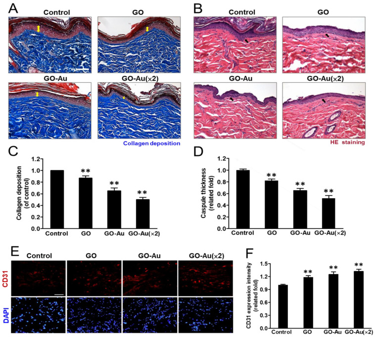
Figure 8.
Histological analysis of foreign-body response (FBR) of different materials following four-week subcutaneous implantation. Photomicrographs showing (A) the Masson’s trichrome-stained histological sections and (B) the hematoxylin and eosin (H&E)-stained histological sections of as-prepared nanocomposites. (C) Quantification of collagen deposition (yellow double arrow in panel A) on the histology examination. (D) Analysis of the FBR was revealed with the capsule thickness (arrows in panel B) based on the histology examination. Scale bar = 100 μm. Data were presented as the mean ± SD (n = 6). ** p < 0.01 as compared with the control group. Immunohistochemical analysis of endothelialization. (E) Immunofluorescent staining of explanted nanocomposites of GO, GO-Au, and GO-Au (×2) with surrounding tissue were stained for endothelialization marker of CD31. Cell nuclei were stained with DAPI (blue). Scale bar = 20 μm. (F) The fluorescence intensity of CD31 was quantified and recorded. Data were presented as the mean ± SD (n = 6). ** p < 0.01 as compared with the control group.

