Figure 4.
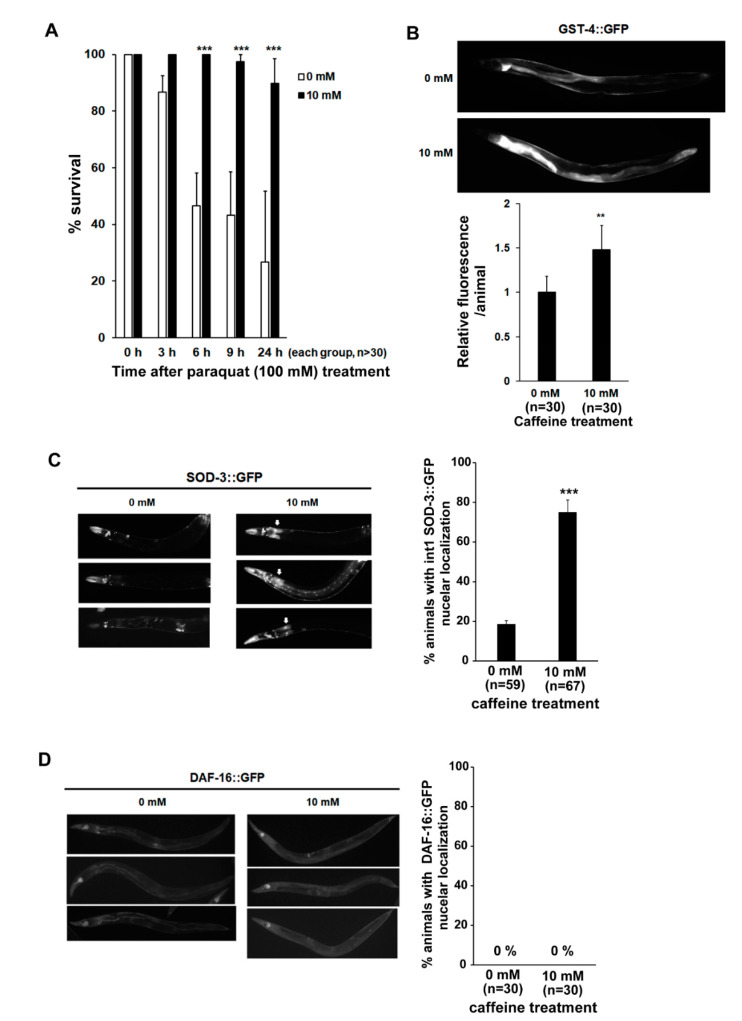
Long-term caffeine intake exerts a protective effect on oxidative stress in advanced ages of C. elegans: (A) Percentage survival rates of caffeine-free diet and caffeine-ingested animals were analyzed under paraquat (100 mM)-induced oxidative stress condition. Error bars represent SD. *** p < 0.001 (two-way ANOVA with Tukey’s post hoc test); (B) Animals expressing glutathione S-transferase 4 (GST-4)::GFP were treated with 0 or 10 mM caffeine at the L4 stage at 25 °C for 72 h. The expression of GST-4::GFP was observed at 72 h post-L4-stage animals at 25 °C. The graph shows the relative fluorescence intensity analyzed by ImageJ. Error bars represent SD. ** p < 0.01 (t-test); (C) Animals expressing superoxide dismutase 3 (SOD-3)::GFP were treated with 0 or 10 mM caffeine at the L4 stage at 25 °C for 72 h. The expression levels of SOD-3::GFP were observed at 72 h post-L4-stage animals at 25 °C. The graph indicates the percentage of animals with SOD-3::GFP nuclear localization in the intestinal cells. Error bars represent SD. *** p < 0.001 (t-test); (D) Animals expressing DAF-16::GFP were treated with 0 or 10 mM caffeine at the L4 stage at 25 °C for 72 h. The expression levels of DAF-16::GFP were observed at 72 h post-L4-stage animals at 25 °C. The graph indicates the percentage of animals with DAF-16::GFP nuclear localization in the intestinal cells.

