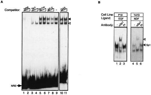
FIG. 4.
Identification of factors interacting with the NRE. (A) Competition between NRE and consensus oligonucleotides. Whole-cell extracts were prepared from NDFβ-treated T47D cells and incubated with the NRE probe in the absence (−) or presence of the indicated unlabeled competitor oligonucleotides at either 100-fold excess (lanes 1, 3, 5, 7, and 10) or 500-fold excess (lanes 2, 4, 6, 8, and 11). An arrow denotes the position of the free NRE probe, and arrowheads denote the positions of the two protein-DNA complexes (the solid arrowhead indicates the slower-migrating complex). (B) The slower-migrating protein-DNA complex is supershifted with an Sp1-specific antibody. Whole-cell extracts prepared from EGF-treated P-19 cells or NDF-treated T47D cells were preincubated with an Sp1-specific antibody (αSp1). As a control, extracts were treated with a preimmune serum (PI) or with no antibody (−). A labeled NRE probe was then added, and protein-DNA complexes were separated on nondenaturing polyacrylamide gels. The positions of the Sp1-specific complex (solid arrowhead) and the supershifted complex (hatched arrowhead) are marked. Note that the lower NRE complex underwent no supershift.

