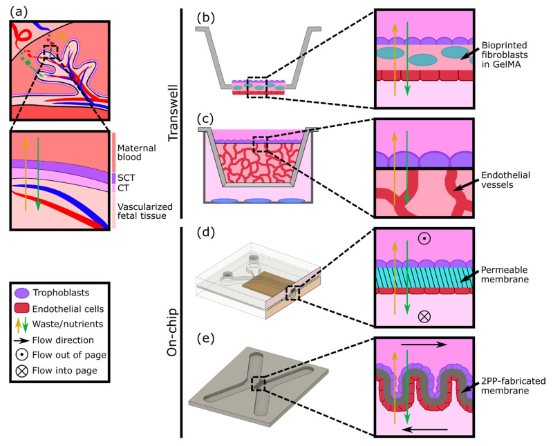
Figure 2.
Modeling the placental barrier in vitro. (a) Diagram of transport between maternal and fetal blood supplies across the syncytiotrophoblast (SCT) and cytotrophoblast (CT). (b) A modified transwell model using a bioprinted layer of fibroblasts with trophoblasts and endothelial cells cultured on either side (based on design in [26]). (c) A transwell model with a layer of trophoblasts cultured on top of vasculature in a 3D gel matrix. Other cell types (blue) can be cultured below the transwell to test the effect of cell secretions (based on design in [27]). (d) Endothelial cells and trophoblasts can be cultured on either side of a permeable membrane in a PDMS microfluidic device with flow (based on designs in [17,28]). (e) Endothelial cells and trophoblasts may also be cultured on either side of a 2PP-fabricated membrane to achieve different geometries (based on design in [29]).

