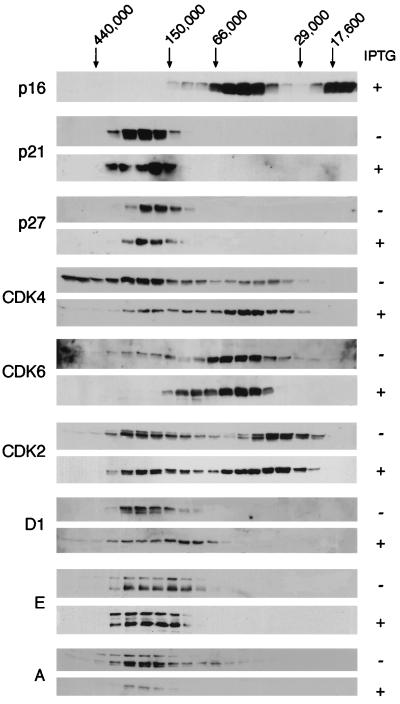
FIG. 4.
Altered size distribution of cyclins, CDKs, and CKI after induction of p16INK4a. Equal amounts (4 mg) of lysate from EH1 cells before (−) and after (+) treatment with IPTG were subjected to gel filtration chromatography. Individual fractions were separated by SDS-PAGE and immunoblotted directly with antibodies against various cell cycle components, as indicated on the left. The elution positions of molecular weight standards are indicated at the top.

