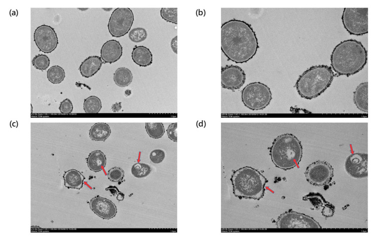
Figure 4.
TEM image of TSP-AB-03 strain untreated or treated with AS101. The morphology of untreated group was observed under a magnification of 12,000× and a scale bar of 1 μm (a) and 20,000× and a scale bar of 1 μm (b). Those treated with 1× MIC (2 μg/mL) of AS101 were captured under a magnification of 12,000× and a scale bar of 1 μm (c) and a magnification of 20,000× and a scale bar of 1 μm (d). The irregular peri-sphere and vacuole inside bacterial cell (red arrows in (c) and (d)) implied the bacterial cell lysis.

