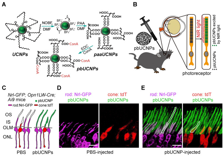
Figure 4.
Photoreceptor-binding upconversion nanoparticles (pbUCNPs) and their distribution in the retina. (A) The surface modification for photoreceptor-binding UCNPs (pbUCNPs) is presented. (B) Schematic diagram to show the subretinal injection and binding of pbUCNPs to the photoreceptor outer segments and green light generation after exposure to near-infrared (NIR) light. (C) Schematic diagram to show pbUCNP (green) distribution in the retina. The cones were labeled with Opn1LW-Cre;Ai9-lsl-tdTomato (pseudo color red), and the rods were labeled with Nrl-GFP (pseudo color violet). (D,E) The mice were injected with PBS (D) and pbUCNP, and the self-anchored pbUCNPs (green) are presented as dashed lines in both the inner and outer segments of rods (violet) and cones (red). The outer nuclear layer is abbreviated as ONL; the outer limiting membrane is abbreviated as OLM; the inner segment of the photoreceptors is abbreviated as IS; the outer segment of the photoreceptors is abbreviated as OS. Reprinted with permission from [15]. Copyright Elsevier, 2019.

