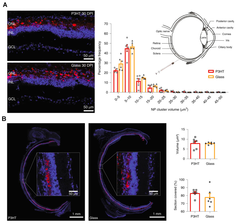
Figure 5.
Wide and stable coverage of subretinally injected P3HT nanoparticles in the retina. (A) The Royal College of Surgeons (RCS) rats were injected with fluorescent glass nanoparticles or P3HT NPs at 30 DPI. The image shows the presence of NP fluorescence (red) with bisbenzimide nuclear staining (blue). Z-stack confocal retinal section images were used to estimate the percentage frequency distribution of the volume of NP clusters at 30 DPI. p > 0.10, two-sided Kolmogorov–Smirnov test. (B) A full equatorial reconstruction of RCS rat retinas injected with the indicated NPs at 30 DPI showed the NP (red) diffusion in the whole subretinal space, with no distribution or penetration in the internal retinal layers (insets at higher magnification). The bar graph shows the total NP volume in the subretinal space and the percentage coverage of retina by NPs at 30 DPI. p > 0.05, two-sided Mann–Whitney U-test. The bar graphs represent mean ± s.e.m. of n = 6 RCS rats for each experimental group. Reprinted with permission from [50]. Copyright 2020 Springer Nature.

