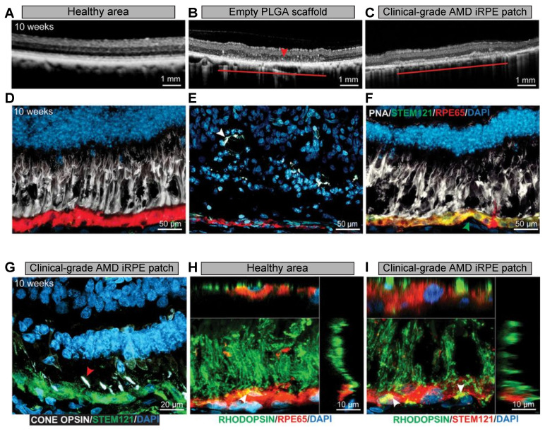
Figure 6.
Integration and functional assessment of AMD-patient-derived iRPE patch in laser-induced porcine retinal degeneration model. (A–C) OCT images of healthy retina and retina transplanted with PLGA scaffold or AMD-iRPE patch. The horizontal line shown in (B,C) indicates the transplanted scaffold over an area of laser-induced RPE ablation. (D–F) The retinal sections were stained with PNA (white), STEM121 (green), and RPE65 (red). The green arrow in (F) indicates the integration of AMD-iRPE (green, STEM121) in the injured porcine retina (red, RPE65). The white arrowhead in (E) shows the presence of retinal tubulations. (G) The retinas were immunostained for red, blue, and green cone opsins (white) and STEM121 (green) to observe the presence of pig cone receptors above human iRPE (red arrow). (H,I) The retinas were immunostained with rhodopsin and RPE65 or STEM121. The white arrow shows the presence of phagocytosed photoreceptor outer segments in both the healthy pig RPE (stained with RPE65) and in human iRPE cells (STEM121). Z-sections show the localization of photoreceptor outer segments. Reprinted with permission from [51]. Copyright The American Association for the Advancement of Science, 2019.

