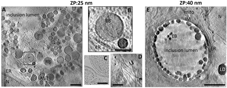
Figure 1.
Observation using cryo-SXT and characterisation of HeLa cells infected by C. trachomatis. HeLa cells infected with C. trachomatis for 24 hpi and observed using cryo-SXT with a 25 nm zone plate (A,B,D) or 40 nm zone plate (E). MiT: mitochondria, N: nucleus, EB: elementary body, RB: reticulate, IM: inclusion membrane, ER: endoplasmic reticulum body, LD: lipid droplets black arrow: inner membrane invagination; (B) corresponds to the box in A and highlights the presence of the pathogen synapse with the T3SS array (white arrow heads); (C): from Dumoux et al. (2012), TEM of the pathogen synapse for reference. D: host cell cytoskeleton. Scale bars: 1 µm (A,D), 0.5 µm (B), 250 nm (C) and 5 µm (E).

