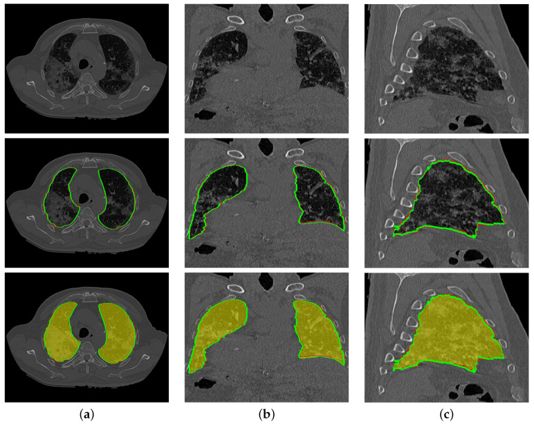
Figure 9.
An illustrative example of the proposed segmentation (second and third rows) for a severe COVID-19 infected lung at (a) 2D axial, (b) coronal, and (c) sagittal cross sections of an original image (first row). Note that red border (green border or yellow region) refers to ground-truth (segmentation).

