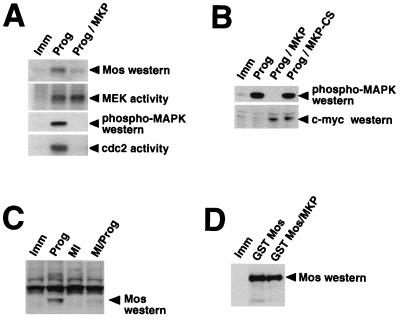
FIG. 1.
MAPK signaling is required for Mos protein accumulation. (A) Immature oocytes were injected with MKP RNA, as indicated, and left for 14 h to express the protein. The oocytes were then stimulated with progesterone (Prog) or left untreated (Imm). Protein lysates were prepared when the progesterone controls reached GVBD50. The same pooled lysate preparations were used for the following analyses: Mos protein accumulation was measured by Western blotting with Mos antisera; the MEK activity assay was performed as described in Materials and Methods with KN-MAPK as substrate; MAPK activation was measured with phospho-MAPK antisera; cdc2 activity assay was measured as described in Materials and Methods with histone H1 as the substrate. (B) Immature oocytes were preinjected with MKP or MKP-CS RNA, as indicated, and left for 14 h to express the protein. The oocytes were stimulated with progesterone (Prog) or left untreated (Imm). Oocyte lysates were used to measure MAPK activation (as described for panel A) and to measure the expression of myc-tagged MKP and MKP-CS by immunoblotting with anti-c-myc antibody. (C) Immature oocytes in medium containing 1% DMSO (MEK inhibitor solvent) were pretreated for 1 h with 100 μM MEK inhibitor (MI) as indicated. The oocytes were then stimulated with progesterone (Prog) or left untreated (Imm). Protein lysates were prepared when progesterone-treated samples reached GVBD50. Mos protein accumulation was analyzed as described for panel A. A nonspecific band, larger than Mos, was also detected by the antibody in all lanes. (D) Immature oocytes were injected with MKP RNA, as indicated, and left for 14 h to express the protein. The oocytes were then injected with RNA encoding GST Mos or left untreated (Imm). Protein lysates were prepared 8 h later, and GST Mos accumulation was measured by Western blotting with Mos antibodies.

