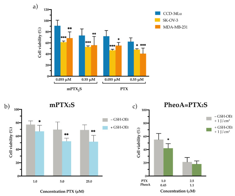
Figure 4.
In vitro cytotoxicity of nanoparticles in different microenvironments. (a) Cell viability measured in normal fibroblasts (CCD-34-Lu) and cancer cells (breast MDA-MB-231 and ovarian SK-OV-3) incubated for 24 h with increasing concentrations of mPTX2S or PTX delivered in the standard solvent and measured 24 h post cell-release in drug-free medium. * p < 0.05; ** p < 0.01; *** p < 0.001 significantly different from CCD-34Lu (ANOVA One-way with Bonferroni’s correction). (b) Cytotoxicity induced by mPTX2S measured in MDA-MB-231 cells pre-treated or not with GSH-OEt (90 min) before nanoparticles incubation (5 h), to further increase the reductive microenvironment in vitro. (c) Cytotoxicity measured in MDA-MB-231 cells (pre-treated or not with GSH-OEt) incubated with PheoA≅PTX2S and irradiated with red light (1 J/cm2). * p < 0.05; ** p < 0.01 significantly different from-GSH-OEt (Student’s t-test). All data in the figure are expressed as mean ± S.D. of at least two independent experiments carried out in triplicate.

