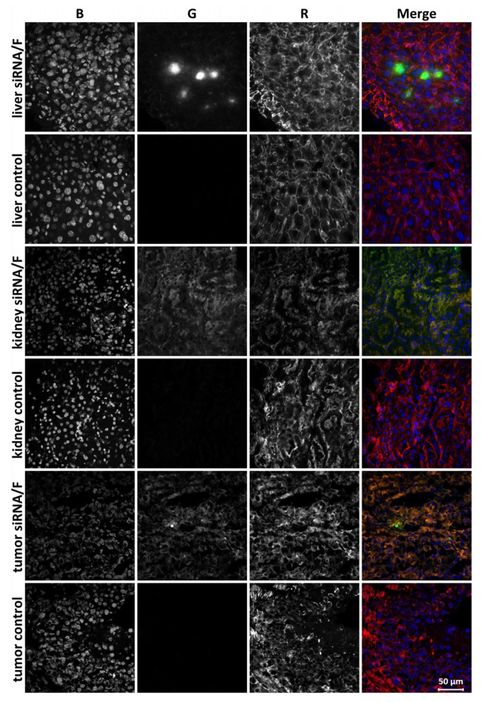
Figure 4.
Accumulation of Alexa Fluor 488–siMDR in the tumor and internal organs of KB-3-1 tumor-bearing SCID mice 24 h after intravenous (IV) injection of Alexa Fluor 488–siMDR/F lipoplexes. Localization of Alexa Fluor 488–siMDR was analyzed by confocal fluorescence microscopy at 200× magnification. Three-channel (RGB) pictures were obtained using Alexa Fluor 488 (R), attached to siMDR; actin filaments are stained by TRITC-phalloidin (G) and DNA is stained with DAPI (B).

