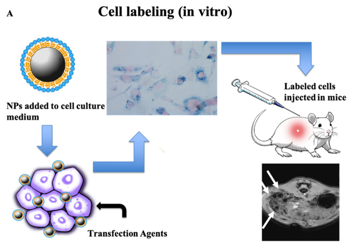
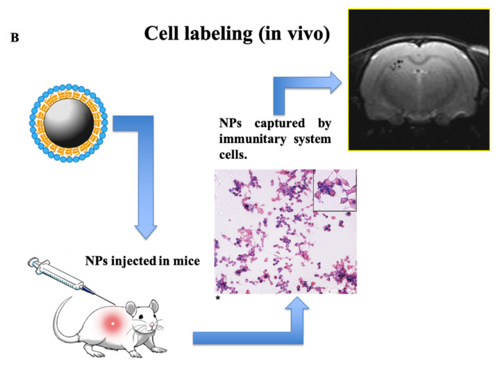
Figure 1.
In vitro and in vivo labeling of cells by using iron oxide NPs. (A) Experimental protocol adopted to label cells in vitro: NPs are added to the cell culture medium, if possible in the presence of transfection agents. After tests on cell viability and iron content, labeled cells are transplanted into living organisms and their in vivo homing can be visualized by MRI. (B) The experimental protocol for in vivo labeling of cells consists in injection of NPs into the bloodstream, where they are captured by immune system cells. MRI therefore allows visualization of regions of immune cell accumulation. The histological image in Figure 1 B(*) is modified from ref. [24].


