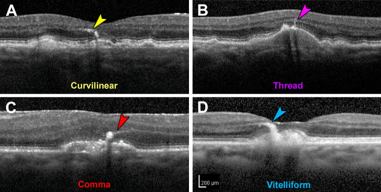
Figure 2.
Morphology of retinal pigment epithelium plumes. Examples from two eyes of one patient. (A) “Curvilinear” consists of hyperreflective structures of consistent width, often arising from a soft druse and aiming toward the outer plexiform layer. (B) “Thread” has a uniform and thin width, compared to curvilinear. (C) “Comma” has a wide base adjacent to the RPE layer and an anterior taper. (D) “Vitelliform” is thick and arises from an acquired vitelliform lesion.24

