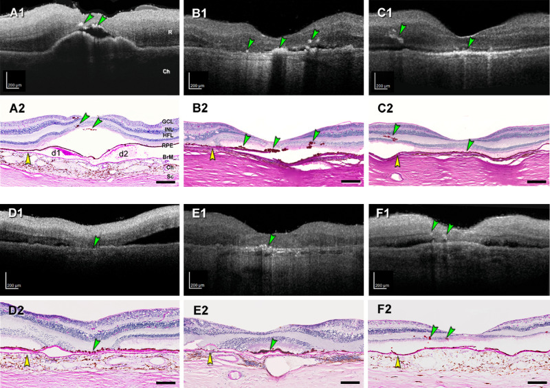
Figure 4.
HRF in ex vivo OCT match intraretinal RPE on histology. Reflective features including HRF are visible in donor eyes (eye numbers in Supplementary Table S7) by ex vivo optical coherence tomography (A1–F1). These were matched with RPE phenotypes in histology sections stained with periodic acid–Schiff hematoxylin (PASH) (A2–F2), to show Bruch's membrane (BrM), basal laminar deposit, and drusen. In all panels, scale bars are 200 µm; yellow arrowheads indicate BrM, and green arrowheads indicate HRF in OCT and ectopic RPE in histology. (A1, A2) HRF above two soft drusen (d1, d2) at fovea from early to intermediate AMD eye (#5). (B1, B2) HRF at the fovea and nasal and temporal side, from atrophic AMD eye (#17). (C1, C2) RPE plume in atrophic AMD eye (#15). (D1, D2) Hyperreflective dots in OCT and dissociated RPE atop thick BLamD in the atrophic area. (E1, E2) Reflective dots under an island of surviving photoreceptors in the fovea center of an atrophic AMD eye (#12). (F1, F2) Multiple HRFs nasal to the fovea in an early to intermediate AMD eye (#7), corresponding to intraretinal RPE cells. Ch, choroid; GCL, ganglion cell layer; INL, inner nuclear layer; R, retina; Sc, sclera.

