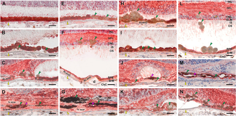
Figure 8.
Abnormal RPE is CRALBP–, and Müller glia are CRALBP+. Twenty donor eyes (16 AMD, 4 controls; eye numbers in Supplementary Table S7) were used for immunohistochemistry with mouse monoclonal anti-human CRALBP (Supplementary Table S1) and red reaction product. Fourteen of 16 previously described morphologic phenotypes of RPE27 were identified (Supplementary Table S2). All scale bars are 20 µm. Yellow arrowheads indicate BrM. Green and fuchsia arrowheads indicate CRALBP– and CRALBP+ abnormal RPE phenotypes, respectively. In panel L, labeled Müller glia are seen at the external limiting membrane (ELM), pericellular baskets in ONL (orange arrowhead), and parallel fibers in HFL. (A) CRALBP+ “uniform” RPE, unremarkable (#1). (B) CRALBP– “shedding” RPE, early to intermediate AMD (#6). (C) CRALBP– “sloughed” RPE, atrophic AMD (#13). (D) Both CRALBP+ and CRALBP– “entombed” RPE nvAMD (#18). (E) CRALBP– “nonuniform” RPE nvAMD (#18). (F) CRALBP– “intraretinal” RPE, early to intermediate AMD eye (#7). (G) Both CRALBP+ and CRALBP– “melanotic” RPE, nvAMD (#18). (H) CRALBP– “severe” RPE, atrophic AMD (#12). (I) CRALBP– “subducted” RPE, atrophic AMD (#12). (J) Both CRALBP+ and CRALBP– “entubulated” RPE, atrophic AMD (#12). (K) CRALBP– RPE at “atrophy without BLamD” area, atrophic AMD (#12). (L) CRALBP– “dissociated” RPE, early to intermediate AMD (#7). (M) Variable CRALBP immunoreactivity in “bilaminar” RPE, nvAMD (#20). (N) Variable CRALBP+ and CRALBP– RPE at “atrophy with BLamD” area, nvAMD (#7).

