Figure 2.
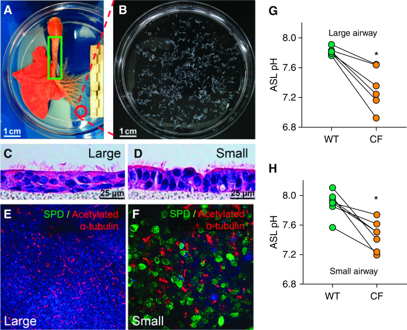
The airway surface liquid (ASL) pH is lower in cystic fibrosis (CF) airway epithelium. (A) The entire airway from the left lung was manually dissected. Sm airways were defined as having a diameter of <200 μm and as lacking cartilage rings and submucosal glands. The green box indicates the region from which Lg airway cells were isolated. Circled region indicates Sm airways, which are shown in B. Scale bars, 1 cm. (C and D) Hematoxylin and eosin staining of well-differentiated, primary, porcine airway epithelial cells. Airway primary cultures were grown on a polycarbonate filter (pore size of 0.4 μm). Note the differentiated epithelium with cilia on the apical surface. Scale bars, 25 μm. (E and F) SPD (surfactant protein D) is detected by immunostaining in cultured primary Sm airway epithelia (F) but not in Lg airway epithelia (E). SPD is stained in green, acetylated α-tubulin is stained in red, and DAPI is shown in blue. Scale bars: E, 100 μm; F, 25 μm. (G and H) The ASL pH was measured using a ratiometric fluorescent dye (SNARF–dextran), which showed that the ASL pH in CF cell cultures from Lg (C) and Sm (D) airways was lower than that in analogous non-CF cell cultures. Each dot represents the measurement taken from a single pig. Data are expressed as the mean ± SE. N = 7 for Sm airway cultures, and N = 6 for Lg airway cultures. *P < 0.05 by Student’s t test. WT = wild type.

