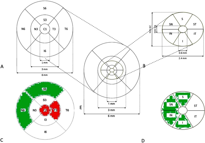
Figure 2.
(A) ETDRS grid centered on the fixation point containing 3 concentric rings with 1, 3, and 6 mm diameters, divided by 2 reticules into 9 subfields: central (C1); inner superior ring (S3), temporal (T3), inferior (I3) and nasal (N3); outer superior ring (S6), temporal (T6), inferior (I6), and nasal (N6). (B) Ganglion cell layer grid (GCL): 14.13 mm2 elliptical annulus area (dimensions: a vertical inner and outer radius of 0.5 and 2.0 mm and a horizontal inner and outer radius of 0.6 and 2.4 mm, respectively) centered on the fovea. Sectors: superior (S); superonasal (SN); inferonasal (IN); inferior (I); inferotemporal (IT); and superotemporal (ST). (C) Comparison of retinal thickness between FEs and CEs in each subfield. If the subfield is thicker in FEs as compared to CEs, it is displayed in red, if thinner in green. If the difference is not statistically significant, it is displayed in white. (D) Comparison of GCL thickness between FEs and CEs in each subfield. If the subfield is thicker in FEs as compared to CEs, it is displayed in red, if thinner in green. If the difference is not statistically significant, it is displayed in white. (E) Overlayed representation of ETDRS and GCL grid.

