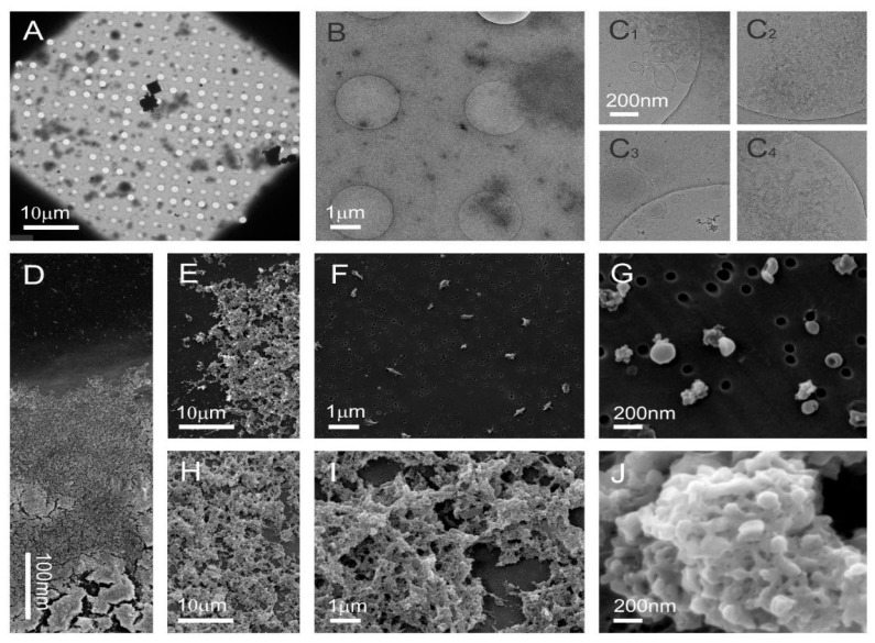
Figure 5.
Cryo-TEM and SEM images of sucrose density separated nanovesicles in the low-density visible Fraction B1 isolated from tomatoes homogenate associated with an SDS–PAGE profile (Figure 3a) that did not show the presence of viral proteins. Cryo-TEM images (A–C) and SEM images (D–J) show the sample rich with sub-micron sized particles that are heterogeneous in size and shape, and numerous membrane-enclosed vesicles.

