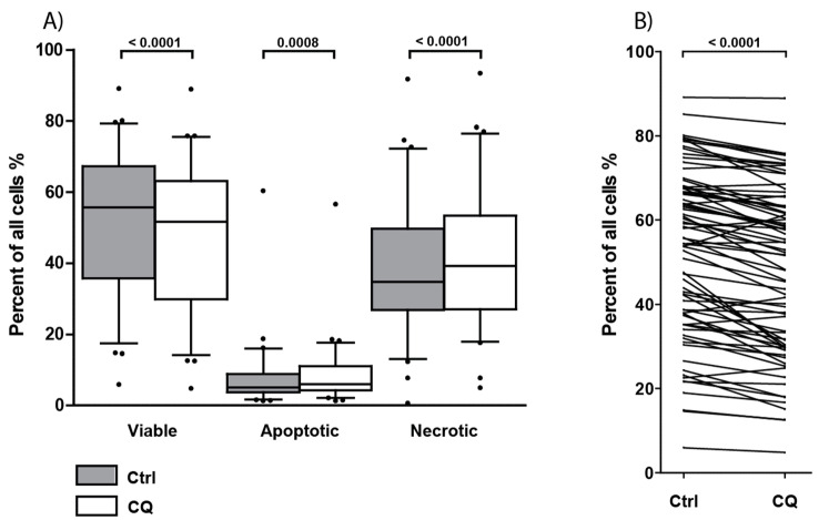
Figure 5.
Effect of chloroquine (CQ) 5 µM on primary AML cell viability, apoptosis, and necrosis after 48 h of culture. Primary cells from 78 consecutive AML patients were cultured with or without 5 µM CQ for 48 h before viability, early apoptosis, and late apoptosis/necrosis were determined by flow cytometry using the AnnexinV/Propidium iodide (PI) assay. (A) The figure shows the overall results for the 72 patients with more than 5% viable cells in drug-free controls. The percentage of viable (AnnexinV− PI−), early apoptotic (AnnexinV+ PI−) and end stage apoptotic and necrotic cells (AnnexinV+ PI+) were determined in patient samples after treatment with 5 µM CQ (white boxes) and after culture in medium alone (gray boxes) for 48 h. Data are presented as median levels, 25/75 percentiles, and 5/95 percentile whiskers, • = outliers. The overall effect when analyzing samples pairwise was examined; and treatment with 5 µM CQ significantly decreased viability (p-value < 0.0001) and increased apoptosis and late apoptosis/necrosis (p-values = 0.0008 and <0.0001, respectively; Wilcoxon signed rank test). (B) The percentage of viable primary AML cells cultured in medium alone compared to treatment with 5 µM CQ for 48 h. This figure presents the viability results for each individual patient. A wide range of cell viability is seen among patients, with a significant decrease in viability after treatment with 5 µM CQ (p-value = 0.0001).

