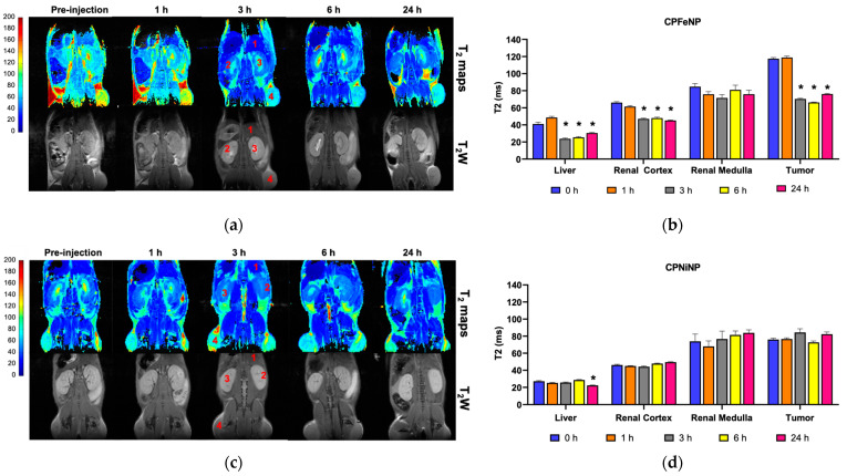
Figure 7.
T2 color-code based maps and T2W images of heterotopic GBM model. T2 color-code based maps and T2W images acquired before the i.v. administration of the IONP-doped CPNs, and at 1, 3, 6 and 24 h after the injection. Numbers indicated the tissues where the ROIs were selected to do the measurements: 1, liver; 2, renal cortex; 3, renal medulla; 4, tumor. (a) CPFeNP MRI evaluation; (c) CPNiNP MRI evaluation. Graphics show the variation in T2 values (mean ± SEM) along the temporal evaluation: (b) CPFeNP; (d) CPNiNP. Statistical symbols correspond to the comparison of the data to the pre-injection value (* p < 0.05).

