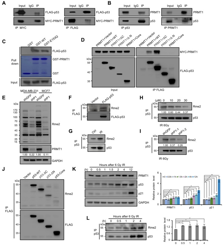
Figure 4.
p53 was methylated on arginine residues in cells. (A) Co-IP assays were performed in HEK293T cells transfected with MYC-PRMT1 and FLAG-p53 plasmids using anti-MYC (left) and anti-FLAG (right) antibodies, and then immunoblotting using anti-FLAG and anti-MYC antibodies. (B) Co-IP assays were performed in MCF7 cells using anti-p53 (left) and anti-PRMT1 (right) antibodies. (C) GST-pulldown assay was performed using FLAG-p53 vector transfected HEK293T cell lysates incubated with GST, GST-fused PRMT1-WT, or -E153Q, followed by Western blotting with anti-FLAG antibody. The protein levels of GST and GST-fused PRMT1 were shown by Coomassie Brilliant Blue staining. (D) Domain mapping of p53 region interaction with PRMT1. FLAG-p53 or its truncations were co-expressed with MYC-PRMT1 in HEK293T cells and precipitated using anti-FLAG beads. Co-IPed PRMT1 was detected by Western blotting using anti-MYC antibody. Left, input. (E) Whole cell-lysates from shPRMT1 and shGFP MDA-MB-231/MCF7 cells were immunoblotted with anti-Rme2 antibody. (F–I) Detection of p53 arginine methylation by Western blotting using anti-Rme2 antibody. (F) FLAG-p53 was immunoprecipitated with anti-FLAG antibody from HEK293T cells expressing FLAG-p53. (G) p53 was immunoprecipitated with anti-p53 antibody from MCF7 cells after 2 h of IR treatment (6 Gy). (H) p53 was precipitated using anti-p53 antibody from MCF7 cells after 2 h of 6 Gy IR; cells were treated with increasing concentrations of Fur (0, 10, 20 or 30 μM) for 24 h. Fur: Furamidine dihydrochloride. (I) p53 was precipitated using anti-p53 antibody from shPRMT1 or shGFP MCF7 cells after 2 h of 6 Gy IR. (J) Domain mapping of the p53 methylation region. FLAG-tagged p53 truncations were expressed in HEK293T cells and precipitated using anti-FLAG magnetic beads. Methylation was analyzed by Western blotting using anti-Rme2 antibody. (K) Detecting the protein levels of PRMT1, p53, and p21 by Western blotting in MCF7 cells after exposure to 6 Gy IR. Quantification shown on the right. Data were presented as mean ± SD from three independent experiments. (L) Western blotting using anti-Rme2 antibody for the detection of the methylated p53 purified from MCF7 cells after 6 Gy IR. The arginine methylation levels were normalized by p53.Quantification shown on the right. Data were presented as mean ± SD from three independent experiments. #, p > 0.05; *, p < 0.05; **, p < 0.01.

