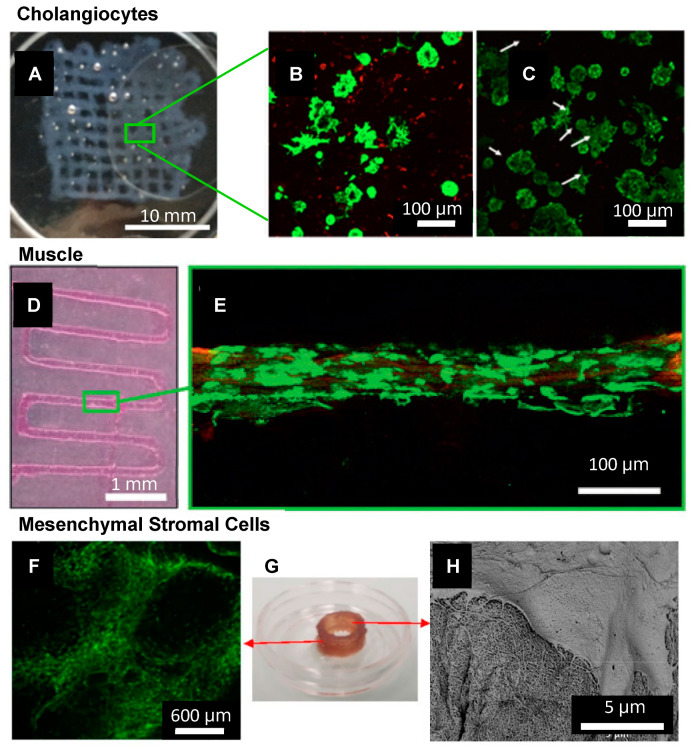
Figure 4.
SAP bioinks have been used for the biofabrication of several cell/tissue types including (A–C) Cholangiocytes. (A) 3D bioprinting IKVAV-ink via extrusion through a 250 μm tip into a 15 mm × 15 mm grid, treated by secondary crosslinking solution. Scale bar is 10 mm. (B) Live/Dead stain of cholangiocytes in IKVAV-ink for 7, and (C) 14 days. Scale bars are 100 μm. Adapted with permission from Yan et al. 2018 Copyright IOPScience [188] (D,E) Muscle. (D) 3D bioprinting of PA-bioink on a CaCl2-coated glass coverslip using a 200 µm nozzle. Scale bar is 1 mm. (E) Confocal image of myoblast cells encapsulated in a filament after seven days in culture, stained with Calcein-AM (green) showing live cells aligned along the fibre axis. Scale bar is 100 μm. Adapted with permission from Sather et al. 2021 Copyright Wiley-VCH [82]. (F–H) MSCs (F) Long-term (30 days) cell viability of hBM-MSCs post-printing of a 1 cm cylindrical construct using IZZK peptide. Scale bar 600 μm. (G) Printed construct after 30 days (H) SEM of printed hBM-MSCs after ten days of culture showing an interaction between the cell’s filopodia and the matrix. Scale bar 5 μm. Adapted with permission from Susapto et al. 2021 Copyright ACS [81].

