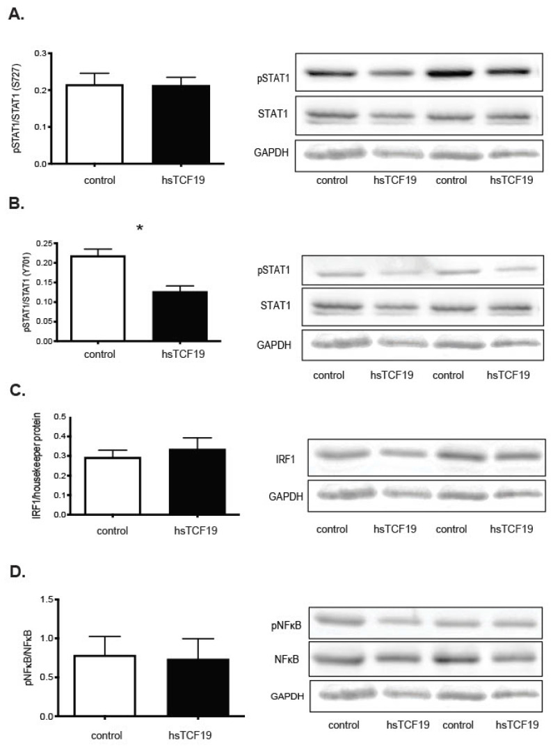
Figure 5.
Overexpression of TCF19 in INS-1 cells does not lead to increased activation of transcription factor targets but leads to decreased activation of STAT1 Y701 phosphorylation. Representative images of two western blot replicates along with analysis of all replicates are shown. Densitometry with Image J 1.44o was used to quantify the bands on the western blots, which were then normalized to the housekeeper protein band. (A) Serine 727 phospho-STAT1/STAT1 protein expression does not show a statistically significant difference between control and hsTCF19 overexpressing cells (n = 5) (phospho-STAT1~91 kDa, STAT1~84, 91 kDa). (B) Tyrosine 701 phospho-STAT1/STAT1 protein expression shows statistically significant decrease in hsTCF19 overexpressing cells compared to control (n = 3) (phospho-STAT1~84, 91 kDa). (C) IRF1 protein levels are not significantly different. Representative western blots in the figure have IRF1 levels normalized to GAPDH. Three other replicates are normalized to beta tubulin (n = 5) (IRF1~48 kDa). (D) There is no difference in phospho NF-κB/NF-κB levels with hsTCF19 overexpression (n = 3) (phospho NF-κB~65 kDa, NF-κB~65 kDa). All data are means ± SEM * p < 0.05.

