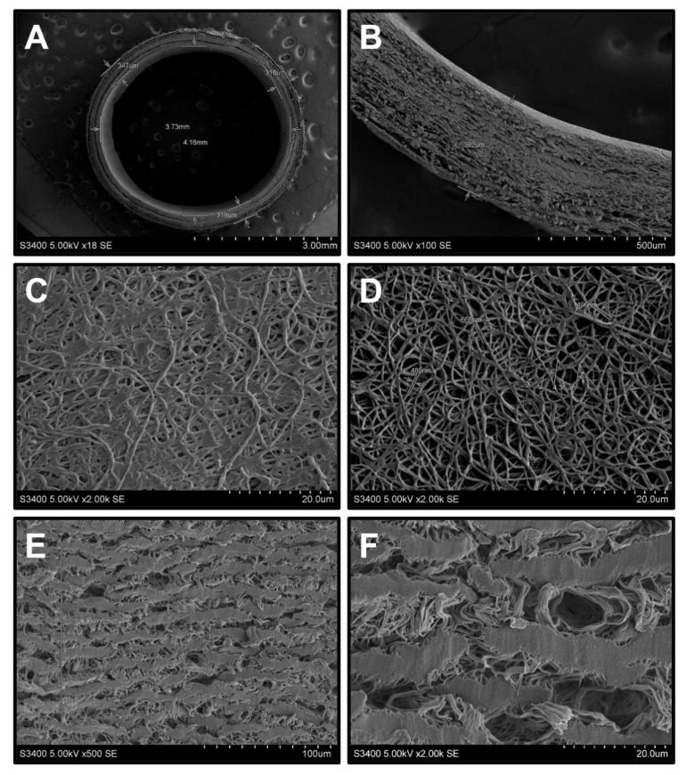
Figure 1.
Scanning electron microscopy of biodegradable and synthetic vascular grafts: (A) Layer-by-layer hierarchical structure of the PHBV/PCL[VEGF-bFGF-SDF]Hep/Ilo grafts, view from above, ×18 magnification; (B) Layer-by-layer hierarchical structure of the PHBV/PCL[VEGF-bFGF-SDF]Hep/Ilo grafts, side view, ×100 magnification; (C) Luminal surface of the PHBV/PCL[VEGF-bFGF-SDF]Hep/Ilo grafts before the washing from PVP, ×2000 magnification; (D) Luminal surface of the PHBV/PCL[VEGF-bFGF-SDF]Hep/Ilo grafts after the washing from PVP, ×2000 magnification; (E) Luminal surface of ePTFE tubular scaffold, ×500 magnification; (F) Luminal surface of ePTFE tubular scaffold, ×2000 magnification. Representative images.

