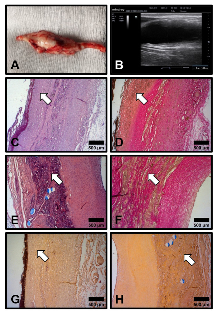
Figure 3.
Development of the aneurysms in the patent PHBV/PCL[VEGF-bFGF-SDF]Hep/Ilo grafts 6 months postimplantation. (A) Gross examination of the aneurysm which developed through the whole length of the graft; (B) Ultrasound examination of the same aneurysm; (C,D) Neointima (indicated by white arrows) demarcated from the degrading polymer scaffold by an organised layer of collagen fibres. Haematoxylin and eosin staining (C) and van Gieson staining (D), ×50 magnification; (E,F) Degrading polymer scaffold is partially substituted by collagen bundles (white arrows) formed de novo. Haematoxylin and eosin staining (E) and van Gieson staining (F), ×50 magnification; (G,H) Absence of calcium deposits both within the neointima (G, white arrow) and polymer scaffold (H, white arrow). Alizarin red S staining, ×50 magnification. Representative images.

