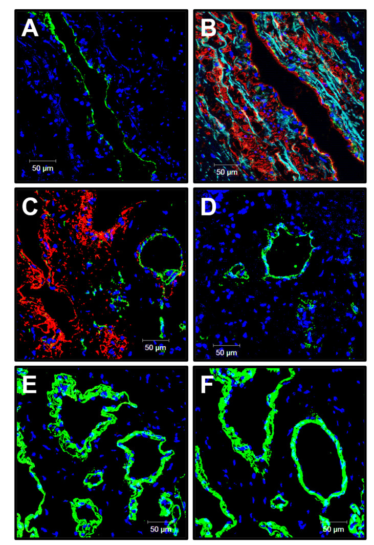
Figure 4.
Confocal microscopy examination of the PHBV/PCL[VEGF-bFGF-SDF]Hep/Ilo graft 6 months postimplantation. (A) CD31 (a marker of endothelial cells, green colour) staining; (B) α-SMA (a marker of VSMCs, red colour) staining; (C) Combined CD31 (green colour) and-SMA (red colour) staining of the vasa vasorum within the graft; (D) vWF (a marker of endothelial cells and a component of the subendothelial ECM, green colour) staining of the vasa vasorum within the graft; (E) Type IV collagen (an ECM protein constituting the vascular basement membrane, green colour) staining; (F) Type III collagen (an ECM protein constituting the vascular basement membrane, green colour) staining. 4′,6-diamidino-2-phenylindole (DAPI) counterstaining (nuclei, blue colour). Representative images, ×200 magnification.

