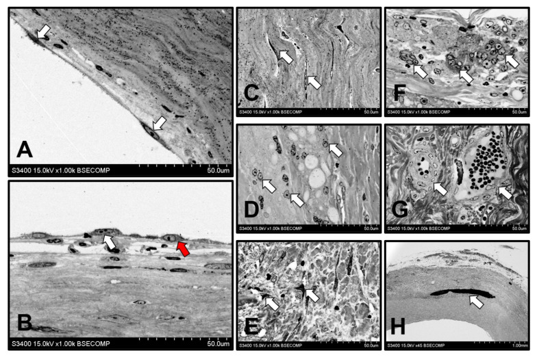
Figure 6.
Backscattered scanning electron microscopy examination of the regenerated artery which replaced PHBV/PCL[VEGF-bFGF-SDF]Hep/Ilo graft 18 months postimplantation. (A) Monolayer of elongated endothelial cells (indicated by white arrows), ×1000 magnification; (B) Combination of elongated endothelial cells (white arrow) and polymorphic endothelial cells (red arrow) suggestive of a transitional phenotype, ×1000 magnification; (C) VSMCs in the neointima (white arrows), ×1000 magnification; (D) Macrophages in the medial layer (white arrows) which substituted a polymer scaffold, ×1000 magnification; (E) Fibroblast-like cells in the medial layer (white arrows), ×1000 magnification; (F) Multinucleated giant cells in the tunica adventitia (white arrows), ×1000 magnification; (G) Adventitial vasa vasorum (white arrows), ×1000 magnification; (H) Calcification on the border of neointimal and medial layers (white arrow), ×45 magnification. Representative images.

