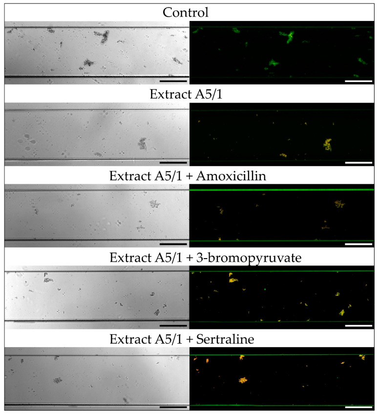
Figure 3.
Representative light and fluorescence microscopy pictures taken at ×10 magnification of H. pylori 8064 strain treated with plant extract A5/1 and its combination with tested synthetic compounds (amoxicillin (AMX), 3-bromopyruvate (3-BP), or sertraline (SER)). Bacteria were treated with substances as follows: MIC of a plant extract, ¼ × MIC of a plant extract + ¼ × MIC of AMX, ¼ × MIC of a plant extract + ¼ × MIC of 3-BP, or ¼ × MIC of a plant extract + ½ × MIC of SER. The study controls were microcapillaries colonized by bacteria not exposed to any antimicrobial substances. In the case of fluorescence pictures, bacteria were stained with the LIVE/DEAD kit, in which green and yellow/orange fluorescence indicates live and damaged/dead bacteria, respectively. Scale bars show 20 µm.

