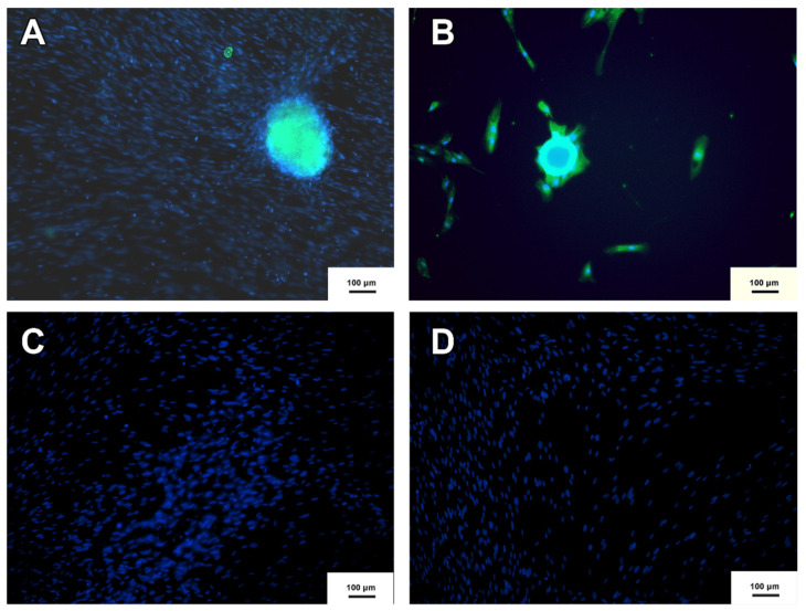
Figure 3.
Neurosphere immunocytochemistry. (A) Neurosphere derived from MSC-WJ (B) Neurosphere derived from MSC-UCB. (C,D) represent the negative controls of both MSC-WJ and MSC-UCB respectively. This means that neither MSC-WJ nor MSC-UCB presented Nestin protein before being seeded in the NFBX. NESTIN protein is shown in green by FITC as a secondary antibody, and Hoechst was used for labeling the nuclei of the cells represented by blue (inverted fluorescence microscope ×100 (inverted fluorescence microscope Axio Vert A1, Carl Zeiss, Oberkochen, Germany).

