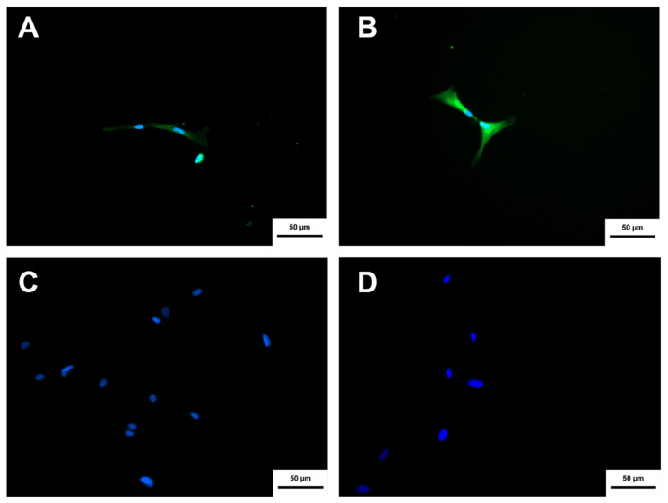
Figure 9.
Cholinergic differentiation immunocytochemistry of NPCN + cells derived from MSC-UCB. (A) MSC-UCB labeling using the anti-ChAT antibody (FITC). (B) MSC-UCB labeling using the anti-ßIII-TUBULIN antibody (FITC). (C,D) represent the negative controls (MSC-UCB not differentiated) for both markers anti-ChAT and anti-ßIII-TUBULIN, respectively Hoechst was used for labeling the nuclei. (Inverted fluorescence microscope with increase of ×100, Axio Vert A1, Carl Zeiss, Oberkochen, Germany).

