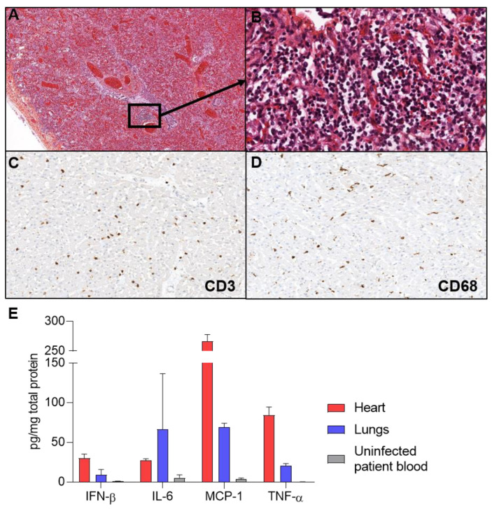
Figure 1.
Pneumonia and myocarditis. (A) Hematoxylin, eosin, and safran-stained sections show scattered foci of necrosis and inflammation (×40) (B) At higher magnification of an inflammatory Figure 400. (C) CD3 immunostaining revealed infiltrates of T lymphocytes in the heart (×400). (D) CD68 immunostaining revealed macrophages cells infiltrates in the heart (×400). (E) Inflammatory cytokines were quantified by ELISA assays, revealing IFN-β, IL-6, MCP-1, and TNF-α increase compared to uninfected patient blood.

