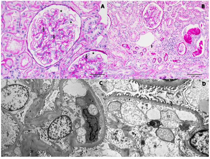
Figure 2.
Light microscopy: (A) Minimal expansion of mesangial matrix (arrows), podocyte hypertrophy (stars) (PAS, 40X). (B) Focal tubular atrophy and interstitial fibrosis (arrow) (PAS 20X). Electron microscopy. (C) Hypertrophic and swollen cytoplasm of severely damaged endothelial cells (EC); the foot processes of podocytes seem to be regularly aligned and inter-digitated. (D) A very mild inner lamina rara widening (arrows) and loss of endothelial cell (EC) fenestrae. A platelet (PT) was also detected in the vessel lumen.

