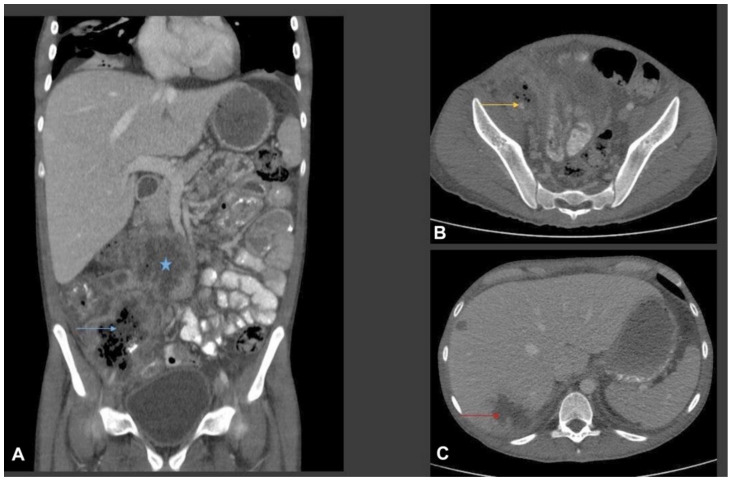
Figure 1.
Multiplanar CT scan of abdomen with oral and intravenous contrast. (A) Coronal CT image shows a multispatial ill-defined low-attenuation ring enhancing collection in the root of the mesentery (star) and in the right iliac fossa. The right iliac fossa collection shows thick irregular rim enhancement with central bubbly air lucency (blue arrow). Bowel loops are adherent to the collections. There is marked inflammatory fat stranding noted in the right iliac fossa and right side of the pelvic cavity. Small volume ascites. (B) Axial CT image from the right iliac fossa and pelvis shows marked phlegmonous soft tissue lesion with fat stranding. The phlegmonous inflammatory mass is encasing the right external iliac artery which is small and irregular in calibre (yellow arrow). (C) Axial images from the liver show multifocal ill-defined low-attenuation liver lesions of variable size (two of them shown in this image). The largest was seen in the subcapsular liver in the segment VII and shows surround oedema, suggestive of liver abscess (red arrow).

