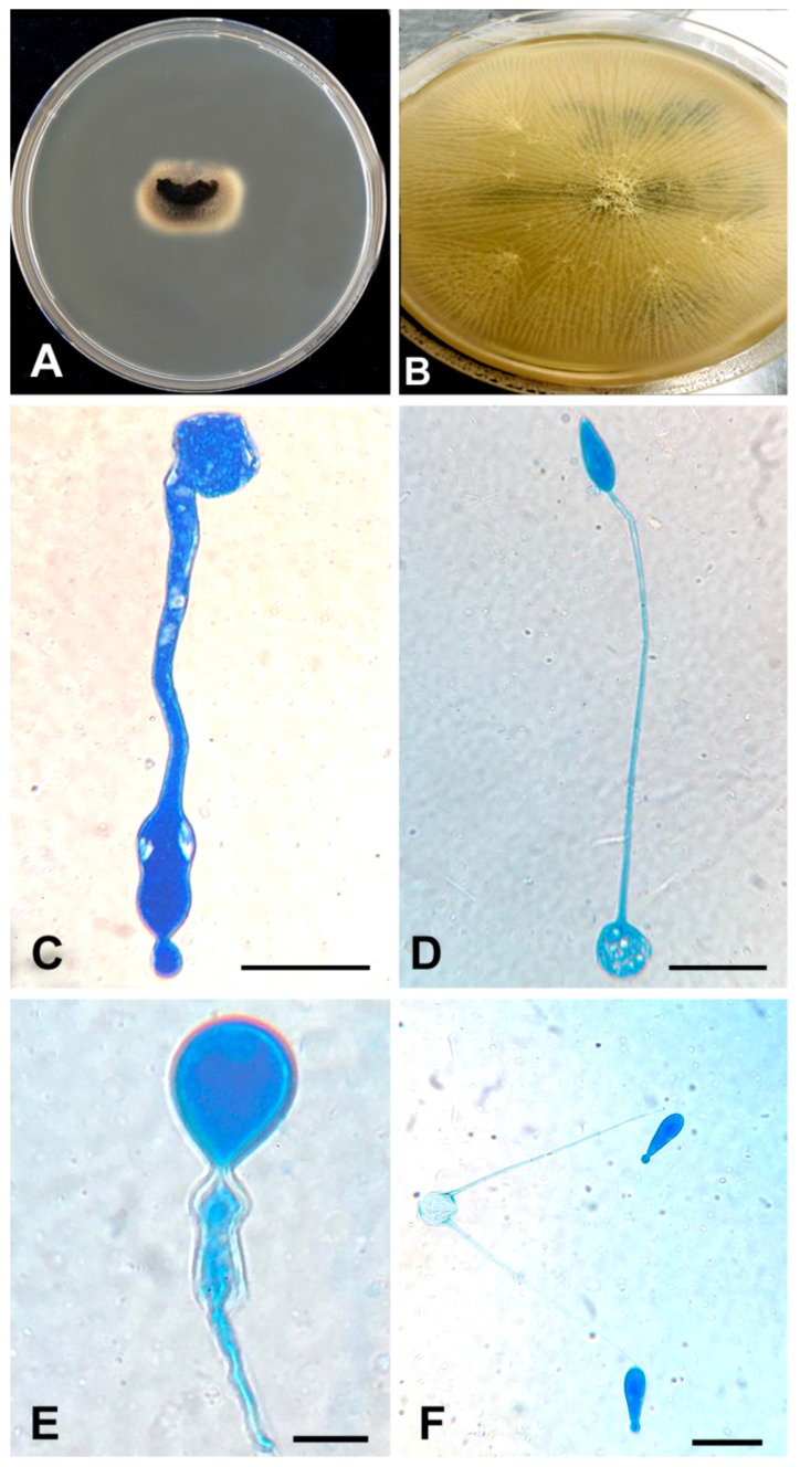
Figure 2.
(A) Direct culture (SAB) from the thrombus. A piece of the thrombus (the black material at the centre) was put on a SAB plate. After incubation, the mould grew from it. (B) Colonial appearance of flat, radially folded, waxy, yellow-cream colonies on SAB. (C–E) Wide hyphae and club-shaped spores with knob-like tips demonstrated with lactophenol cotton blue stain. (F) The thin ballistoconidia in the primary culture. All scale 10 µm.

