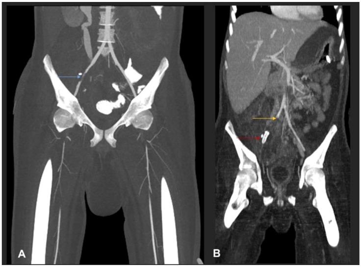
Figure 4.
(A) MIP coronal CT angiography of aorta and lower limb shows a segmental narrowing and luminal irregularities of right external iliac artery (blue arrow). No occlusion. Distal run off was satisfactory. Rest of the lower limb vessels were patent and normal (not shown). (B) MIP coronal image of the aorta and lower limbs shows cut-off of the proximal right common iliac artery suggestive of thrombotic occlusion (yellow arrow). A stent is seen in the right external iliac artery (red arrow). Right iliac fossa shows extensive inflammatory phlegmonous mass-like soft tissue thickening.

