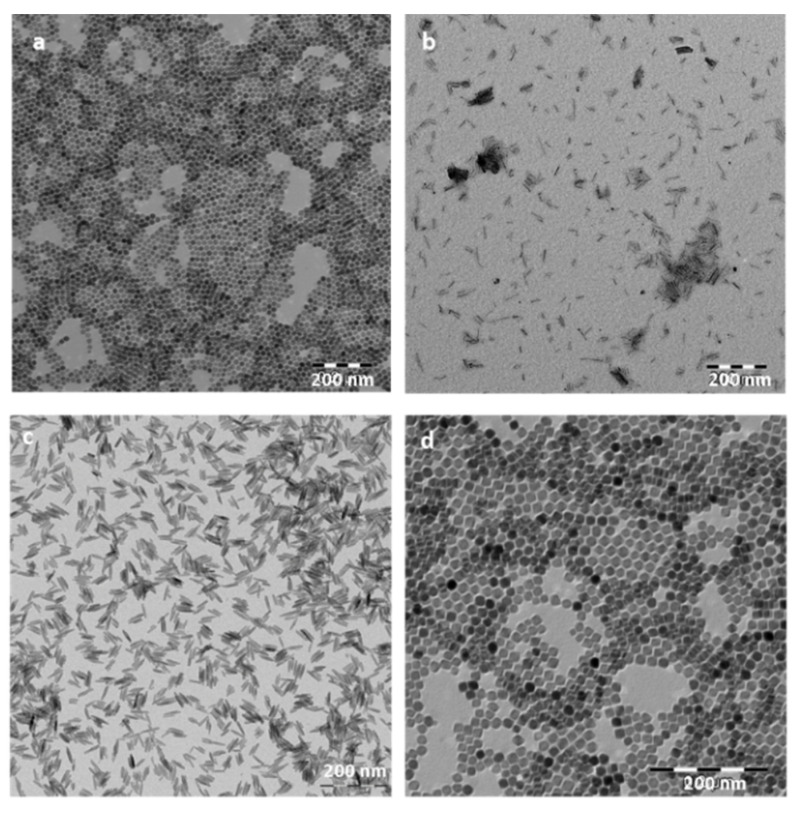
Figure 5.
Transmission electron microscopy (TEM) images of the iron oxide nanoparticles (magnetite) prepared by the hydrothermal method with different morphologies: (a) spheres, (b) rods, (c) needles, and (d) cuboidals. Images were obtained using a LIBRA 120 Plus Carl Zeiss microscope (A Carl Zeiss SMT AG Company, Oberkochen, Germany).

